The Vanderbilt University Institute of Imaging Science (VUIIS) offers several noninvasive imaging resources through the Human Imaging Core (HIC) to provide both standard and advanced functional, structural, and quantitative imaging of the human visual system. Protocol design and image processing resources are also available.
Resources
- Magnetic Resonance Imaging (MRI)
– Three research-dedicated MR scanners are available for use: two 3 Tesla scanners and a 7 Tesla scanner.
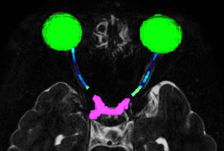
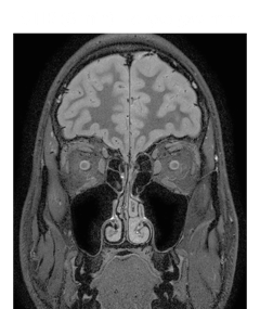
– A wide variety of MR imaging techniques are available at both 3 and 7 Tesla to investigate the human visual system, including (but not limited to) imaging of optic nerve myelination, high resolution structural imaging of the thalamic nuclei, and functional imaging (fMRI) of the visual cortex. - Near Infrared (NIR) Optical Tomography
– Oxyhemoglobin, deoxyhemoglobin and total hemoglobin concentration changes can be recorded in response to visual stimuli.
– NIR optical tomography is an alternative to fMRI for patient populations that cannot tolerate MRI. - Optical Coherence Tomography (OCT)
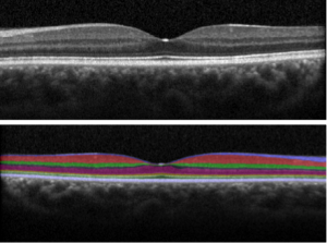
– Near infrared light is used to visualize and measure the thickness of the retinal layers.
– Applications include detection and monitoring of diseases such as glaucoma, diabetes, multiple sclerosis, and Alzheimer’s.
– Quality assessment reports are provided for each scan.
Human Imaging Core FAQs
- Where is the VUIIS Human Imaging Core?
– Vanderbilt University Hospital (VUH): 3Ta
– VUH B160 (In the basement across from the in-patient pharmacy)– VUIIS: 3Tb, 7T, NIRS, and OCT
– 1161 21st Ave S, Nashville, TN 37232 (Enter from Medical Center Drive) - Who can use the VUIIS imaging resources?
– The VUIIS is a trans-institutional core serving VU and VUMC physicians, scientists, and trainees, as well as corporate affiliates. - How do I get started with a project the VUIIS?
– A new Project Request must be submitted and approved for each imaging resource you intend to use for your study.
– To access the VUIIS HIC resource calendars for scheduling, you and your staff will need to:
– Register with the VUMC core management system (click HERE for instructions)
– Request access to the VUMC Institute of Imaging Science (VUIIS) – Human Imaging
core within the core management system (you must be logged in to do this)
Hardware and Software at the 3T
Patrick Henry is primarily responsible for the hardware used for stimulus presentation and behavioral data collection at the 3T scanner. Specifically, he is responsible for the proper functioning of the Mac and PC computers and monitors for stimulus presentation, and for the audio-visual presentation systems (e.g. projectors, screens, goggles, head phones), and non-imaging-related data collection (eye tracker). Pat is also responsible for installation and maintenance of commonly used software on these computers, including Matlab, e-prime, and of software that drives peripherals (e.g. eye tracker). He is not responsible, however, for other equipment or peripherals at the scanner (e.g. head coils). If the fMRI technologist on duty cannot troubleshoot the latter equipment, he/she may contact appropriate Phillips personel. Pat is available to trouble-shoot any of the hardware and software issues mentioned above. He can be reached either via telephone (office: 322-4584), pager (831-4602) or e-mail. Pat’s office in Wilson Hall is at 529WH. In addition, to better serve the fMRI community, Pat will also have a second office at the computer lab of the Vanderbilt University Institute of Imaging Science (VUIIS), adjacent to the scanner suite (room B163). Please keep in mind that Pat may not be able to arrive immediately to solve problems, and that the problem may not be easily or quickly solvable. In the rare event when Pat is not available, you or the fMRI technologist may contact Ken Wilkens (ken.wilkens@vanderbilt.edu, phone: 2-4019), Senior VUIIS Engineer, who will try his best to help out.
Software Installation
In the past, we have had a policy that any users can install new software on the computers at the scanner, but only after making an announcement to the fMRI community. This policy has served us well, and we will therefore continue with it, but please remember to announce any software change to the fMRI community (using the fmri@list.vanderbilt.edu email list). It would be useful, however, if we could keep track of the location of the installation software, in case some programs need to be re-installed at short notice. Thus, if you have installed software on one of the console room computers, please contact Pat to let him know. Pat is also available to help in installation of fMRI-related software and hardware in individual labs, including the transfer, import, and reading of fMRI data. At this time, Pat is not able to teach individual laboratories how to use particular fMRI-related software (e.g. SPM, BV), or advise you in data-analysis issues. Similarly, please address problems regarding fMRI data integrity (e.g. spikes) to VUIIS members (e.g. Chris Gatenby, Ken Wilkens). If you are uncertain where to direct specific fMRI issues or requests, Pat will provide assistance in locating the appropriate resources. Finally, in addition to his functions at the 3T MRI scanner, Pat also ensures proper functioning of the TMS system and optical imaging systems in Wilson Hall.