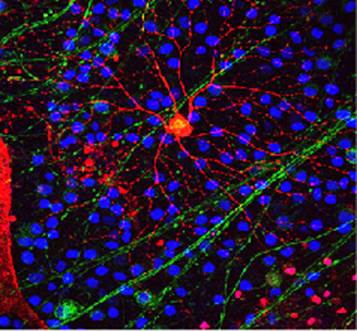
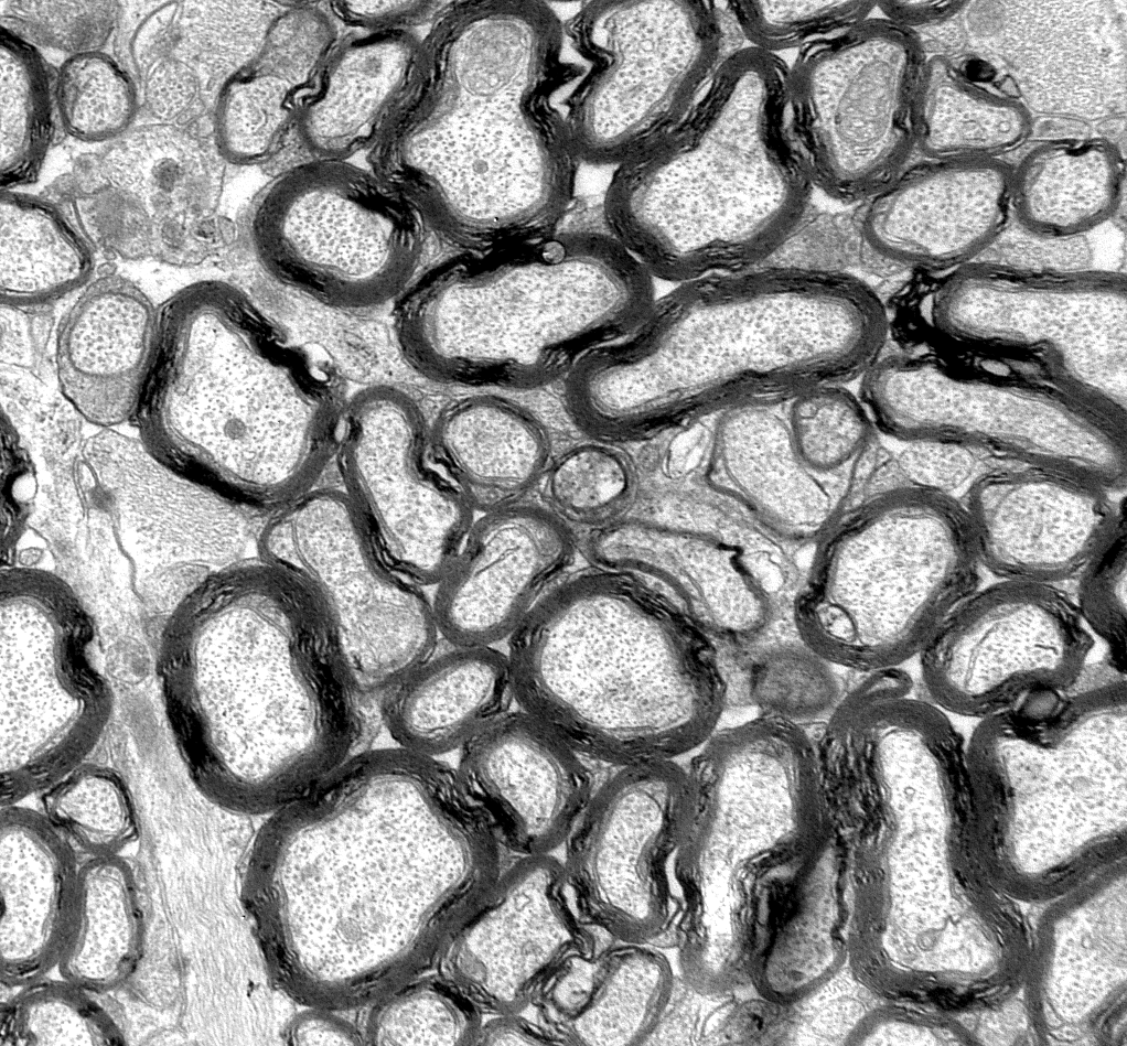
The Cell Imaging and Analysis Core offers a variety of microscopy-based imaging platforms via the Vanderbilt Cell Imaging Shared Resource (CISR). This resource is a multi-center core facility providing state-of-the art microscopic imaging technologies. These include an array of specialized confocal, laser‑scanning, and high-resolution microscopes as well as conventional optical microscopes. The core also provides high-performance computational facilities for image processing. Investigators wishing to use the confocal microscope schedule time through an on-line calendar maintained by the CISR. Information regarding access and equipment scheduling is posted on the CISR web site. Information includes (a) descriptions of the facility and equipment, (b) a form for registration and account setup, (c) instructions for operating the equipment, (d) contact information for CISR staff, and (e) a mechanism for self-scheduling (by trained users) of all microscope systems. An initial contact usually involves a brief discussion of intent and sample description to define the appropriate equipment requirements, then a training session or feasibility experiment is scheduled. The general operating policy of the CISR requires all new and infrequent users be trained and demonstrate basic competency prior to gaining unsupervised access to the microscopes. Following demonstration of competency, scheduling access is enabled and equipment is available 24 hours per day, 7 days per week.

