The PROTEIN ANALYSIS SERVICE will be housed in the Mass Spectrometry and Proteomics Core Facility of the Mass Spectrometry Research Center (MSRC) which occupies 3000 sq ft on the 9th floor of Medical Research Building III (see map in Appendix I) with space for mass spectrometry instruments, protein separation instrumentation, computing support and laboratory personnel. This space is immediately adjacent to laboratories of the Caprioli (Director of the MSRC) and Hachey (Director of the Proteomics Core Facility) and Schey (Director of VVRC Protein Analysis Service) research groups, which will facilitate interactions with Mass Spectrometry and Proteomics Core personnel. Located in Medical Research Building III places them in the center of the NEI funded investigators on campus. The Core houses PC based workstations for mass spectrometry instruments and sample processing robotics workstations, including a dedicated PC workstation for 2D gel image analysis and bioinformatics. The Proteomics Core has 7 Pentium class computers in offices and labs for general laboratory and office functions. There are 13 Pentium class computers dedicated to instrument control and robotics applications, 2 data workstations and one 9 node Sequest PVM bioinformatics workstation. All of the local MSRC instruments are on a private network, but have access through a network firewall to the campus network. The Vanderbilt Mass Spectrometry and Proteomics Core is outfitted with state of the art instrumentation for protein and peptide analytical separations and mass spectrometry analysis as described below.
- Three ThermoFisher LTQ linear quadrupole ion trap LC MS MS systems. These quadrupole ion trap instruments are used for LC MS MS analyses and are equipped with nano electrospray sources, autosamplers and quaternary LC pump systems to deliver nanoliter flow rates for high sensitivity analyses. The linear trap technology exceeds performance of widely used 3D traps in both sensitivity and scan speed, which enables detection of a greater variety of peptides in complex mixtures, including a greater number of low abundance and modified forms. The instruments are equipped with Xcalibur and Bioworks 3.3 software. Related equipment includes an Agilent 1100 series LC workstation for ion exchange peptide separations for multidimensional LC MS MS analyses and equipment for fabrication of micro LC columns.
- One ThermoFisher LTQ Orbitrap (LC/MS) ion trap MS. This newest generation in ion trapping technology combines the advantages of the LTQ instruments described above with the high resolution and high mass accuracy of the Orbitrap mass spectrometer. These advantages provide for more accurate assignment of peptide sequences and more accurate determination of protein modifications. This instrument is equipped with an Eksigent nanoLC Plus HPLC workstation that delivers nanoliter/min flows via a splitless pumping system.
- Applied Biosystems Voyager 4700. This instrument combines a MALDI source with a TOF TOF tandem mass analyzer to acquire both peptide mass fingerprint data and MS MS spectra of peptide ions via high energy collision induced dissociation. The instrument is equipped with Explorer 4700 instrument control software and GPS Explorer 2.0 software suite for proteomics data analyses. Unique automation capabilities of the ABI 4700 provide for high throughput analyses and enable LC MALDI MS MS analyses via off line collection of LC fractionated peptide mixtures. This approach complements electrospray LC MS MS for analysis of complex protein and peptide mixtures.
- ETTAN DIGE 2D gel electrophoresis system (Amersham Biosciences). Includes equipment for first dimension isoelectric focusing (Multiphor II and IPGphor) and second dimension SDS PAGE (Ettan DALT II) for large format (25 x 20 cm) gels. Protein samples are individually labeled with sensitive (sub picogram) cyanine fluorescent dyes (Cy2, Cy3 or Cy5) that exhibit a linear dynamic range of approximately four orders of magnitude. Additional instrumentation includes Amersham Ettan Spot Picker, Digester and MALDI plate spotter, which permit high throughput, automated analyses of protein features selected from 2D gels.
- Two Leica cryostats (CM 3050S and CM 3600) are used for sectioning frozen tissues for MALDI profiling/imaging. The CM 3600 is a large cryostat that can be used for whole animal sectioning.
- Zeiss Mirax slide scanner is used to acquire high resolution digital images of tissue sections on slides. The scanner is used for histology directed studies that relate mass spectrometry generated protein profile to morphological features of tissue sections.
- Two Labcyte Portrait 630 reagent multispotters are state of the art high resolution automated liquid spotting systems. Using a patented acoustic spotting mechanism ultralow, accurate spotting of any liquid can be applied to tissue sections for subsequent mass spectrometric imaging. These devices offer rapid, reproducible, accurate spotting of samples with high spatial resolution.
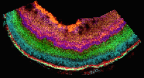
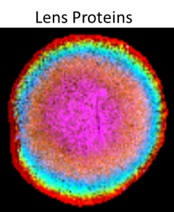
- Bruker AutoFlex II reflector time of flight mass spectrometer is dedicated to MALDI tissue imaging experiments and offers a high speed (200 Hz) laser with Smartbeam technology for improved performance and reflector technology for enhanced mass resolution. Equipped with FlexImaging software, acquisition of MALDI tissue images is highly automated in a user friendly environment.
- ThermoFisher MALDI Duo LTQ linear ion trap mass spectrometer is used for small molecule (drugs/metabolites, etc) imaging from tissues. This high sensitivity mass spectrometer combined with the MALDI source provides a powerful combination for small molecule imaging including the ability to utilize tandem mass spectrometry for highly specific studies of targeted molecules.

